Category: Bài báo ngoài nước

  • Giới thiệu công bố khoa học

    Giới thiệu công bố khoa học

    Tiêu đề bài báo: Granulocyte colony-stimulating factor reduces biliary fibrosis and ductular reaction in a mouse model of chronic cholestasis

    Tạp chí: Liver Research, số tháng 3 năm 2023

    Toàn văn: https://www.sciencedirect.com/science/article/pii/S2542568423000053

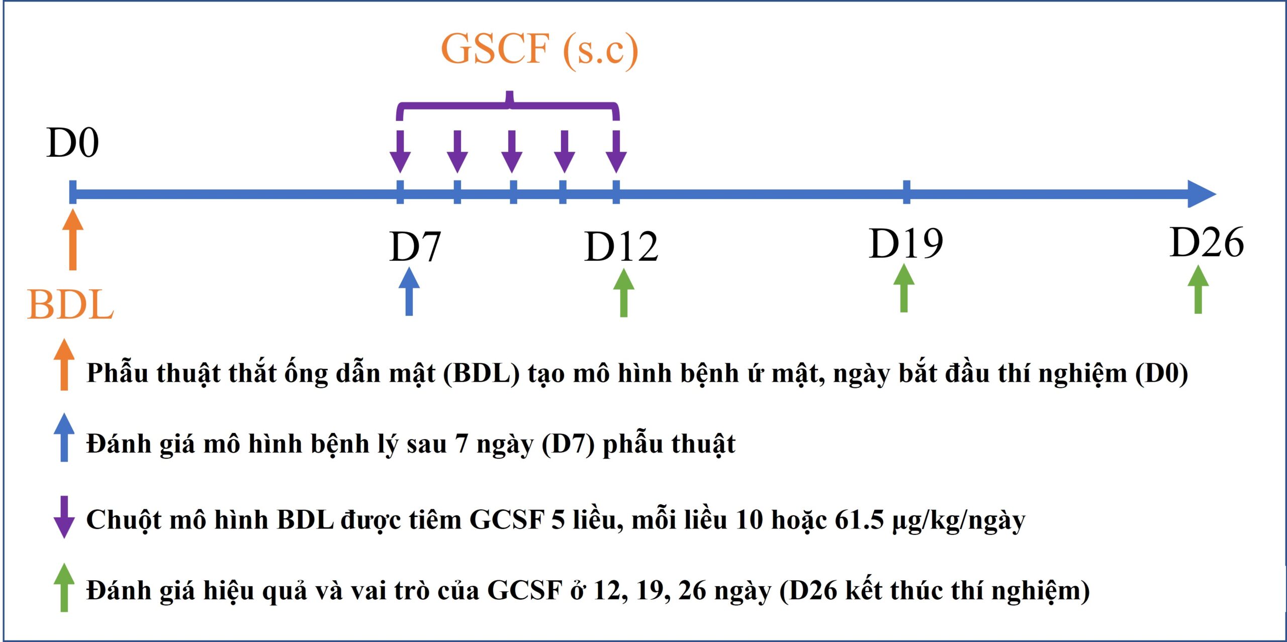
    Nhân tố kích thích bạch cầu hạt (GCSF) là protein của cơ thể tác động lên tế bào gốc tủy xương tăng sinh, phân chia và di cư. Đây là thuốc được chỉ định điều trị thường quy cho các bệnh nhân bị suy bạch cầu ở người. Đồng thời, GCSF củng được ứng dụng trong việc tăng cường huy động tế bào gốc ra ngoài máu ngoại vi để thu nhận và cấy ghép cho bệnh nhân ở nhiều nghiên cứu khác nhau. Trong nghiên cứu này, nhóm tác giả đã sử dụng GCSF để ứng dụng điều trị mô hình chuột bị bệnh lý gan do tắc nghẽ đường mật, mô hình mô phỏng bệnh lý hẹp đường mật ở người.

    Hiện nay, hẹp đường mật là một bệnh lý có tình trạng diễn tiến nhanh với tỷ lệ tử vong và tái phát cao sau điều trị đặt ra yêu cầu về việc phát triển liệu pháp mới giúp hỗ trợ điều trị, kéo dài thời gian sống cho bệnh nhân là rất cần thiết. GCSF đã được chứng minh có nhiều vai trò giúp hỗ trợ điều trị trên các bệnh lý gan giai đoạn cuối như tăng cường sự di cư của tế bào gốc về mô tổn thương, kích thích sự tăng sinh tế bào gan và điều hòa đáp ứng miễn dịch. Do đó, nhóm nghiên cứu tái tạo gan (Hepatoregeneration) tiến hành nghiên cứu về hiệu quả và vai trò của GCSF trên mô hình bệnh lý gan mạn tính do ứ mật.

    Kết quả của công bố cho thấy, hiệu quả điều trị bệnh gan mạn tính do ứ mật của GCSF phụ thuộc vào liều lượng thuốc sử dụng. Đồng thời, nghiên cứu cũng chỉ ra các cơ chế tiềm năng của GCSF trong điều trị bệnh gan trên mô hình tắc nghẽn đường mật này gồm (i) tăng cường huy động tế bào gốc di cư về gan; (ii) kích thích sự tăng sinh tế bào gan; (iii) ức chế tế bào gây xơ là nguyên bào sợi cửa; and (iv) ức chế phản ứng ống mật. Các kết quả này là cơ sở cho việc ứng dụng GCSF trên lâm sàng trong điều trị bệnh lý hẹp đường mật.

    Hepatoregeneration, PTN Nghiên cứu và Ứng dụng Tế bào gốc, Trường đại học Khoa học Tự nhiên, Đại học Quốc gia Hồ Chí Minh.
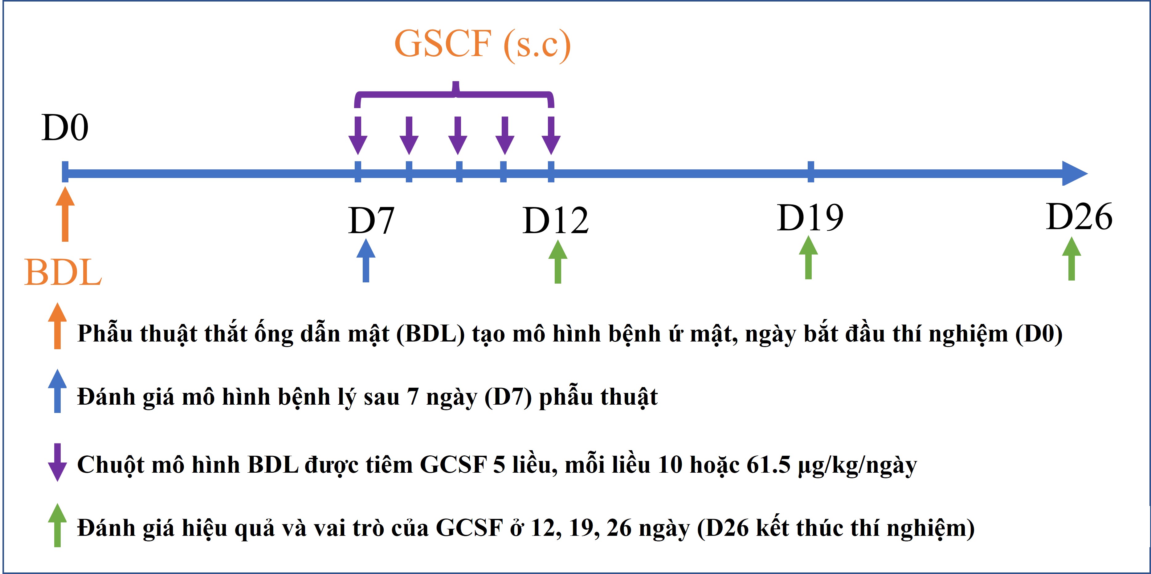
    Sơ đồ mô tả nghiên cứu
  • Transplantation of umbilical cord blood-derived mesenchymal stem cells to treat liver cirrhosis in mice: a comparison of tail and portal vein injection

    Abstract

    Introduction: To date, there have been many studies indicating the positive effects of stem cells on treating liver cirrhosis. In this study, we used umbilical cord blood-derived mesenchymal stem cells (UCB-MSCs) for treatment in a mouse model of liver cirrhosis. Specifically, we determined and compared the effectiveness of two methods of MSC injection (tail vein versus portal vein).

    Methods: Liver cirrhosis in male Swiss mice (of age approximately 11 weeks or under) was induced by administration of carbon tetrachloride (CCl4; 1 ml/kg). One million UCB-MSCs were then transplanted into cirrhotic mice via the portal vein or tail vein. After 21 days, blood samples were collected for measurement of transaminase, bilirubin and albumin. The expression of fibrosis-associated genes, specifically procollagen – alpha 1 and integrin – beta1, were assessed using quantitative RT-PCR. The histopathology of the specimens was also evaluated using hematoxylin/eosin, Masson trichrome staining, and immunohistochemistry using collagen type 1 and alpha-SMA antibodies.

    Results: After 21 days, cirrhotic mice treated with UCB-MSCs showed recovery of bilirubin index, increase of liver albumin synthesis, inhibition of fibrosis-related gene expression (e.g. procollagen – alpha 1 and integrin – beta1), and remodeling of liver histology. From comparison of the different routes of transplantation, UCB-portal route was significantly more effective than UCB-tail route at reducing aspartate transaminase (AST) activity and bilirubin index (P<0.05), and inhibiting procollagen – alpha 1 and integrin – beta1 expression (P<0.05). UCB-MSCs from both transfusion routes showed accelerated improvement of liver histopathology.

    Conclusion: Therapeutic strategies using UCB-MSCs have proven to be promising for the treatment of liver cirrhosis. Injection of UCB-MSC via portal vein was more effective than tail vein for cirrhosis treatment.

    Ngày công bố: 09/09/2017
    Link: http://www.cellstemcell.org/index.php/PSC/article/view/365

  • Comparative treatment efficiency of adipose and bone marrow derived allogenic mesenchymal stem cell transplantation in mouse models of liver fibrosis

    Abstract

    Background: The application of mesenchymal stem cell (MSC) therapy in liver fibrosis treatment has been increasingly investigated in recent years. MSCs obtained from a variety of sources (e.g. bone marrow, umbilical cord blood and adipose tissue) have been studied and have achieved remarkable results. In this study, we compared the effects of adipose-derived mesenchymal stem cells (AD-MSC) transplantation with bone marrow-derived mesenchymal stem cell (BM-MSC) transplantation in a mouse model of liver fibrosis, induced by carbon tetrachloride (CCl4).

    Methods: Eight-week old mice were treated with CCl4 for 11 weeks to induce liver fibrosis then 5×10⁵ cells were transplanted into mice via the tail vein.

    Results: After 21 days of transplantation, the results showed that the stem cell treated groups ameliorated better than the placebo group. MSC treated groups showed reduced AST and ALT levels, down-regulated expression of extracellular matrix (ECM) genes, and improved liver histopathology. Both sources of MSCs (bone marrow and adipose tissue) were effective in the mouse model of liver fibrosis.

    Conclusion: Our results also indicated that AD-MSC transplantation in mice accelerated liver regeneration better than BM-MSC transplantation.

    Ngày đăng: 26/06/2017
    Link: http://www.bmrat.org/index.php/BMRAT/article/view/179

  • Allogeneic Adipose-Derived Mesenchymal Stem Cell Transplantation Enhances the Expression of Angiogenic Factors in a Mouse Acute Hindlimb Ischemic Model

    Abstract

    Cell migration and molecular mechanisms during healing of damaged vascular or muscle tissues are emerging fields of interest worldwide. The study herein focuses on evaluating the role of allogenic adipose-derived mesenchymal stem cells (ADMSCs) in restoring damaged tissues. Using a hindlimb ischemic mouse model, ADMSC-mediated induction of cell migration and gene expression related to myocyte regeneration and angiogenesis were evaluated. ADMSCs were labeled with GFP (ADMSC-GFP). The proximal end of the femoral blood vessel of mice (over 6 months of age) are ligated at two positions then cut between the two ties. Hindlimb ischemic mice were randomly divided into two groups: Group I (n = 30) which was injected with PBS (100 μL) and Group II (n = 30) which was transplanted with ADMSC-GFP (106 cells/100 μL PBS) at the rectus femoris muscle. The migration of ADMSC-GFP in hindlimb was analyzed by UV-Vis system. The expression of genes related to angiogenesis and muscle tissue repair was quantified by real-time RT-PCR. The results showed that ADMSCs existed in the grafted hindlimb for 7 days. Grafted cells migrated to other damaged areas such as thigh and heel. In both groups the ischemic hindlimb showed an increased expression of several angiogenic genes, including Flt-1Flk-1, and Ang-2. In particular, the expression of Ang-2 and myogenic-related gene MyoD was significantly increased in the ADMSC-treated group compared to the PBS-treated (control) group; the expression increased at day 28 compared to day 3. The other factors, such as VE-CadherinHGFCD31Myf5, and TGF-β, were also more highly expressed in the ADMSC-treated group than in the control group. Thus, grafted ADMSCs were able to migrate to other areas in the injured hindlimb, persist for approximately 7 days, and have a significantly positive impact on stimulating expression of myogenic- and angiogenesis-related genes.

    Ngày đăng: 16/06/2017
    Link: https://link.springer.com/chapter/10.1007%2F5584_2017_63

  • ETV-2 activated proliferation of endothelial cells and attenuated acute hindlimb ischemia in mice

    Abstract

    Ischemia is the reduction of blood flow to tissues by injury of blood vessels. Depending on the sites of tissues and grade of ischemia, ischemia can cause many serious complications. This study aimed to evaluate the effects of the E-twenty six (ETS) factor Ets variant 2 (ETV2) gene expression in angiogenesis and the effect of ETV2 gene therapy in a mouse model of hindlimb ischemia. The role of ETV2 on endothelial cell proliferation was evaluated in vitro. Knockdown of ETV2 expression was done using short hairpin RNA (shRNA) lentiviral viral particles. The ETV2 viral vector was injected into the skeletal muscles at the ligated and burned sites of the hindlimb and evaluated for its efficacy as a gene therapy modality for ischemia. Vascular regeneration in mice was indirectly evaluated by changes in mouse survival, necrotic grades of the leg, normal blood oxygen saturation level (SpO2), and blood flow by trypan blue injection assay. Preliminary data showed that ETV2 expression played a role in angiogenesis of endothelial cells. ETV2 overexpression could trigger and stimulate proliferation of skeletal endothelial cells. In vivo knockdown of ETV2 expression inhibited the auto-recovery of ischemic hindlimb, while overexpression of ETV2 helped to rescue leg loss and reduce necrosis, significantly improving angiogenesis in hindlimb ischemia. Our findings demonstrate that ETV2 gene therapy is a potentially effective modality for vascular regeneration.

    Ngày đăng: 19/04/2017
    Link: https://link.springer.com/article/10.1007%2Fs11626-017-0151-4

  • Extracellular vesicles of ETV2 transfected fibroblasts stimulate endothelial cells and improve neovascularization in a murine model of hindlimb ischemia

    Abstract

    Ischemia are common conditions related to lack of blood supply to tissues. Depending on the ischemic sites, ischemia can cause different diseases, such as hindlimb ischemia, heart infarction and stroke. This study aims to evaluate how extracellular vesicles (EVs) derived from ETV2 transfected fibroblasts affect endothelial cell proliferation and neovascularization in a murine model of hindlimb ischemia. Human fibroblasts were isolated and cultured under standard conditions and expanded to the 3th passage before use in experiments. Human fibroblasts were transduced with a viral vector containing the ETV2 gene. Transduced cells were selected by puromycin treatment. These cells were further cultured for collection of EVs, which were isolated from culture supernatant. Following co-culture with endothelial cells, EVs were evaluated for their effect on endothelial cell proliferation and were directly injected into ischemic tissues of a murine model of hindlimb ischemia. The results showed that EVs could induce endothelial cell proliferation in vitro and improved neovascularization in a murine model of hindlimb ischemia. Our results suggest that EVs derived from ETV2-transfected fibroblasts can be promising non-cellular products for the regeneration of blood vessels.

    Ngày đăng: 04/04/2017
    Link: https://link.springer.com/article/10.1007%2Fs10616-017-0095-2

  • Improving stem cell engraftment to enhance functional efficacy in cardiovascular disease: where are we now?

    Abstract

    Stem cell therapy is a promising therapy for repairing damaged tissue. A growing body of research shows that stem cells work effectively in several diseases such as cardiovascular disease, hepatic disease, and diabetes. It has been shown that stem cells not only differentiate into functional cells and replace dead cells, but also release growth factors and cytokines which can recruit autologous cells. The most significant barrier to achieve clinical relevance of this treatment mode is the poor survival rate of injected cells. To improve transplantation and enhance functional outcome, investigations of gene transfection (overexpression of anti-apoptotic and antioxidant proteins), growth factor supplementation, and scaffolding matrices are being conducted. In this review, we will focus on methods to increase cell survival in stem cell transplantation as a novel treatment for cardiovascular disease.

    Ngày đăng: 31/01/2017
    Link: http://www.bmrat.org/index.php/BMRAT/article/view/146

  • Mesenchymal stem cells for diabetes mellitus treatment: new advances

    Abstract

    Mesenchymal stem cells (MSCs) are the most widely used stem cells of the human body due to ease of successful isolation and expansion for many years. In particular, from 2012 until now, MSCs have been widely clinically used to treat various diseases, including graft versus host disease (GVHD), Crohn’s disease, and knee osteoarthritis. In this review, the applications of MSCs in diabetes will be reviewed and discussed. Diabetes mellitus type 1, also known as Type 1 diabetes (T1DM), is an autoimmune disease in which immune cells attack the beta cells in islets of Langerhans (pancreatic islets). Although type 2 diabetes (T2DM) is considered to be a disease related to insulin resistance, several recent studies have shown some relation of immune dysfunction in this disease. Therefore, MSC transplantation may be a beneficial treatment for both T1DM and T2DM. MSC transplantation in preclinical trials and clinical trials for T1DM and T2DM have shown a moderate to significant improvement in diabetes without adverse side effects. In this review, we will discuss some of the updates from preclinical and clinical trials of MSC transplantation for diabetes.

    Ngày public: 31/01/2017
    Link: http://www.bmrat.org/index.php/BMRAT/article/view/144

  • Adipose tissue derived stromal vascular fraction transplantation can recover spinal cord injury in mice

    Abstract

    Introduction: Stem cell therapy is one of the most promising therapies for degenerative diseases and related injuries. Adipose tissue derived stem cells (ADSCs) exhibit some particular properties such as high production of paracrine factors. Indeed, ADSCs have been successfully used to treat diseases, including osteoarthritis, diabetic ulcer, etc.

    Methods: In this study, ADSCs were used to treat spinal cord injury (SCI) in a mouse model. Non-expanded ADSCs, from stromal vascular fractions (SVFs) isolated from both autologous and allogeneic adipose tissues, were injected into injured sites of mice at a specified dose. The SCI mouse model were generated by transection of spinal cord at vertebrae T8 – T10. After 1 week of transection, mice exhibiting completed SCI were divided into 4 groups: group 1 was control (mice without any treatment), group 2 was placebo (mice treated with platelet rich plasma (PRP)), group 3 was allogeneic SVF transplantation (mice treated with allogeneic SVFs), and group 4 was autologous SVF transplantation (mice treated with autologous SVFs). For the treatment groups, mice were transplanted with 20 µL of activated PRP or/and with 106 cells of SVF (allogeneic or autologous) into the injured position through laminectomy. The recovery of SCI was evaluated by locomotor test, sensory test and sensory-motor test at 5 weeks after transplantation. The histology of the spinal cord also was checked after 5 weeks.

    Results: The results showed that in all groups with PRP injected with or without SVFs, the inflammation was efficiently controlled. The glial scar as well as myelin defragmentation were clearly reduced. However, a significant improvement of BBB score was only recorded in mice transplanted with autologous SVFs.

    Conclusion: The results of our study show that autologous SVF transplantation in combination with PRP can be a promising therapy for SCI.

    Năm: 2016
    Link: http://www.cellstemcell.org/index.php/PSC/article/view/148

  • Autologous and allogeneic transplantation of adipose derived stem cells have similar efficacy for type 1 diabetes mellitus therapy in mouse models

    Abstract

    Introduction: Type 1 diabetes mellitus (T1D) disease is caused by lesions or dysfunction of beta cells of pancreatic islets, causing less insulin to be secreted into the blood and thereby increasing glucose levels in the blood. In this study, we evaluated and compared the efficiency of treatment for T1D using autograft and allograft adipose-derived stem cells (ADSCs). Methods: ADSCs were collected from the belly of mice before they were injected using a single dose of streptozotocin (100 mg/kg) to induce T1D. T1D mice were intravenously injected with a dose of 2×10⁶ ADSCs into the tail vein. Therapeutic efficacy was assessed by survival rate, blood glucose levels, serum insulin levels, histology and immunohistochemistry of pancreatic islets. Results: The results showed that both autograft and allograft transplantation of ADSCs demonstrated similarities in mortality rate, blood glucose level, blood insulin level, quantity and size of pancreatic islets. Both transplantations significantly improved T1D mice, which showed a decrease in mortality rate as well as blood glucose level, and increases in blood insulin level, quantity and size of pancreatic islets. Conclusion: The similar results suggest that both autologous and allogeneic transplantations of ADSCs are promising therapy for T1D treatment.

    Năm: 2016
    Link: http://www.cellstemcell.org/index.php/PSC/article/view/142